After breast diagnostic exams are performed, if there is a mass or an area of concern, a biopsy may be needed to determine if it is benign or cancerous.
During a breast biopsy, a small sample of tissue is obtained using a hollow needle under imaging guidance, such as ultrasound, MRI, or mammography (stereotactic biopsy). Local anesthesia is always used, so the procedure is generally not painful, and is easily performed as an outpatient at the Partners Imaging Women’s Centers at either our Sarasota or Bradenton centers.
The tissue is sent to a pathologist who examines it under a microscope to determine a diagnosis. Your referring physician will share the results with you.
The radiologist will also evaluate the results of the biopsy to make sure that the pathology and imaging findings match up appropriately (concordant). If the results show cancer cells, the surgeon can use this information to plan surgery.
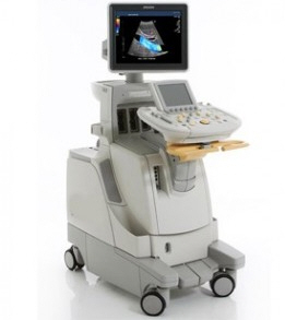 Biopsy can be performed using ultrasound to localize the mass. An ultrasound transducer is placed on the breast as the patient lays on a table, and is used to visualize the area of concern while the radiologist performs the biopsy in real time. We also perform cyst aspirations under ultrasound guidance using the same technique.
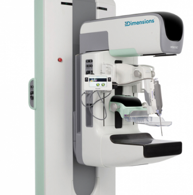
Biopsy can also be performed under stereotactic guidance. Partners has the latest Hologic Affirm upright biopsy system that provides superior imaging and accuracy compared to the traditional stereotactic table.
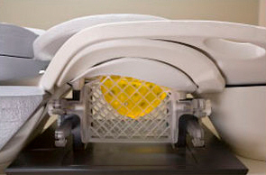
Biopsy can be performed in MRI, using a guided biopsy system that is built into the MRI (see picture below). The area of interest in the breast is localized in 3 dimensions using a grid system. The yellow area represents the breast and how it would look for the technician that performs the scan.
 Each of these methods achieve the same goal- to obtain a sample of tissue from the area of concern so that it can be checked by a pathologist. The radiologist will decide which method to use based on how the abnormality is best visualized.
Usually, calcifications or areas of asymmetry are biopsied using stereotactic guidance, and masses are biopsied using ultrasound guidance. MRI biopsies are usually performed on those abnormalities that are only seen on an MRI scan. Image guided biopsy is a nonsurgical, minimally invasive method of assessing a breast abnormality.
Very little recovery time is needed, and patients can soon resume their usual activities. The procedure is less invasive than surgical biopsy, leaves little or no scarring, and can be performed in less than an hour. Compared with open surgical biopsy, the procedure is about one-third the cost.
Many imaging centers simply perform mammography screening and then rely on other centers or a hospital to carry out the diagnostic work and / or biopsy. This means multiple visits to different facilities, and having records move from one place to another, with a high probability of different radiologists being involved. None of this is necessary with Partners Imaging Women’s Centers. Our nurse navigator will guide you through the entire process.